This tab will serve as a place where I answer my leading questions listed in the about tab and where I post general information on my research topic.
Topic Question: How does adolescent alcohol use affect their brain’s function and structure?
How does adolescent alcohol use affect the user’s brain anatomically or physiologically is often a question research by neuroscientists and psychiatrists. But the question is not often considered by adolescents themselves and their parents. So the goal of this project is to inform and educate teenagers and their parents on the dangers of adolescent alcohol use. The topic id a very prevalent problem here in the United States and around the world, yet only the medical professions seems to have subsequent knowledge on the topic. Everyone is affected by underage alcohol use, whether one uses alcohol while being underage of if they knew someone killed because of it. Before one would learn about how the developing brain’ structure and function were affected by alcohol, one would have to understand the developing healthy brain.
The brain is still developing well into a person’s twenties and the since the brain is the body’s most complex organ, adolescent brain development is the body’s most important process. Teenage brain development is a very simple process with two stages, a growth spurt and the pruning and chunking of neural connections. The growth spurt does not occur during the teenage years but rather during utero and in the first months of life. During this time the brain grows at a rapid pace and produces millions of brain cells. The second part of brain development during adolescence occurs when a person is ten to thirteen years old and it is when the brain organizes and cleans out neural pathways that the brain no longer uses. For example if a teenager is involved in sports and academics, the brain keeps the neural connections used in sports and academics. But if that same person is not involved in music, the brain cleans out the neural connections used in music. This is why people tend to be good at a hobby that they have been doing for an extended period of time. The brain makes room for the neural connections most used by the brain. The pruning process makes way so our brain can become much more efficient and faster. This process also makes room for a long chain of complex nerve cells required for complex problem solving used during childhood (Tapert et al., 2000).
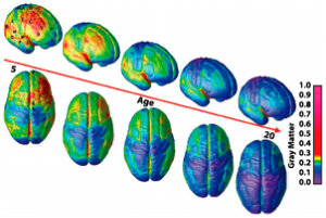
Brain development occurs from front to back, so the pre frontal cortex which is responsible for complex reasoning does not develop well into a person’s twenties. Since the pre frontal cortex is still developing into the teenage years, teenagers may make impulsive decisions. During the teenage years white matter or nerve tissue along with the hippocampus are still developing so all developing areas are going to be more sensitive to alcohol’s effects (Spear et al., 2000).
Above is a picture showing the phases of brain development from the age of five until the age of twenty (Fredrikson et al., 2009).
Guidelines that define my research:
In order to fully understand how alcohol affects adolescent brain anatomy and physiology, one would also need to have some guidelines that outline the key aspects of my research. Adolescents are teenagers from the ages of twelve to twenty. Adolescent alcohol use is defined as any alcohol use because it is illegal but scientists and public health officials seems to think two drinks during adolescence can affect brain function in the long run (niaaa.nih.gov et al., 2014). Because the brain is still developing during adolescence, the effects of alcohol use even in small amounts and only a couple of times can have profound effects on the adolescent brain.
Common terms used in research:
Some keywords that come up while researching how alcohol affects the teenage brain are terms related to the structure and areas of the brain affected by alcohol. Areas mentioned in the research that I have come across are white matter, pre frontal lobes, pre frontal cortex, the hippocampus, physiological functioning, cognitive functioning, visuospatial functioning, the corticles, neurotransmitters, frontal lobes, parietal regions, cerebellar areas, the cerebral cortex, and neuropeptides.
To fully understand how a healthy developing brain’s anatomy is affected by alcohol one must understand the function of a healthy brain and its components. The brain is defined as “an organ of soft nervous tissue contained in the skull of vertebrates, functioning as the coordinating center of sensation and intellectual and nervous activity” (Merriam Webster Dictionary). The brain is composed of four lobes, the brain stem, the cerebellum, the hypothalamus, the amygdala, the corpus colossus, the cerebrum, and the hippocampus. The four lobes are the occipital, parietal, frontal, temporal and the material that makes up the outermost surfaces of the brain are called the cerebral cortex, the motor cortex, the pre frontal cortex, the visual cortex, and the cerebellar cortex. These cortexes cover the surface of their corresponding lobes. The parietal lobe is located in the cerebral hemisphere. This lobe controls comprehension. Visual functions, language, reading, internal stimuli, and tactile sensation along with sensory comprehension are monitored in these lobes. Located within the parietal lobe are the sensory cortex and the motor cortex. They are responsible for monitoring the body’s movement. The frontal lobe is another lobe located in the cerebral hemisphere and it controls creative thought, problem solving, intellect, judgment, muscle movement, personality, and coordinated movement. The third lobe also located in the cerebral hemisphere, is the temporal lobe and it controls visual and auditory memories such as when you see or hear a cat and know it is a cat from visual and auditory memory. The last lobe, the occipital lobe, controls one’s vision. The white matter is the tissue of the nervous system that covers the entire nervous system and the cerebrum; it consists of myelinated nerve axons that transmit signals from one side of the cerebrum to the other and to other areas of the brain and body.
The cerebrum, the largest portion of the brain is responsible for the brain’s overall function. It is divided into the four lobes. The cerebrum contains a left and right hemisphere which is connected by axons that relay messages. The cerebellum is responsible for balance, posture, and coordination and it is often referred to as the little brain.
The limbic systems help relay emotions and it is responsible for hormonal responses. It is composed of the amygdala, the hippocampus, the hypothalamus, and the thalamus. The amygdala responds to emotion, memories, and fear. The hippocampus is responsible for learning memory and making short term memories into long term memories. The hypothalamus controls thirst, hunger, and one’s body temperature and the thalamus controls the attention span.
The brain stem contains the medulla, the pons, and the midbrain. The brain stem is responsible for the functions of basic life such as heart beat, blood pressure, and breathing. The midbrain controls body movement, vision, and hearing and the pons controls heart rate and breathing (Medicine.net et al., 2014).
Because alcohol essentially affects the brains functions, one would also need to know about the different types of brain functioning. There is neuropsychological functioning, physiological functioning, cognitive functioning, and visuospatial functioning. Neuropsychological functioning is brain function that is concerned with relationships between brain and behavior. It includes behavioral and cognitive changes. Physiological functioning focuses on the functioning of the nervous system. Cognitive functioning is a branch of brain functioning that is connected to the function of memory, perception, and association. Lastly visuospatial functioning allows us to visually perceive objects and the relationships among images and objects (Clark et al., 2008).
Another set of terms commonly used in research dealing with alcohol and the developing brain are terms connected to physiological functioning such as neurotransmitters and neuropeptide systems. Neurotransmitters are chemical messengers that transmit the signals throughout the body that control thought processes, behavior, and emotion and are released by the brain at synapses when an action potential releases them. The neurotransmitters than attach themselves to receptor molecules on the synapse’s specific target cell. Neuropeptides are protein molecules used by neurons to communicate with each other. The neuropeptides are signaling molecules that alter brain functions such as analgesia (a deadening of the sense of pain, reward, metabolism, and reproduction.) Neuropeptides function as neuronal communication by acting on cell surface receptors. They act as specific signals between one population of neurons.
Overall dangers of adolescent alcohol use:
Since the adolescent brain is still developing the brain is more vulnerable to some of the effects of substances. Alcohol ingestion during adolescence can have long lasting detrimental effects on normal brain functioning during adulthood. The brain’s key areas are still maturing during the teenage years and are more sensitive to the toxic effects of drugs and alcohol. Immature brain regions may place teenagers at an elevated risk to the effects of alcohol. Since the developing brain is extra sensitive to alcohol’s affects, the brain reduces social discomfort which would cause a more pleasurable experience when a teenager drinks, which makes drinking addictive (Black et al., 2003). Higher metabolic rates in adolescents allow them to consume more alcohol that is safe without feeling sick. Because adolescence is such a critical phase in brain development that alcohol has a greater impact on the adolescent brain than the adult brain. Insults to the brain during adolescence could have an impact on long term brain function (White et al., 2013).
In the adolescent brain alcohol increases the effects of dopamine (the brain’s reward system) by causing more dopamine and dopamine receptors to be created, so the brain is tricked by alcohol into thinking alcohol is a reward. Because of this, the body and brain continues to give it more “rewards” and that is when binge drinking causes adolescents to become sick without noticing. Alcohol suppresses the amygdala which diminishes the psychological experience of stress which makes it easy to participate in risky behaviors. Everyone knows that the typical effects of alcohol on the adult brain, the brain stem shuts down, their balance and coordination are impaired and more often than not people black out, slur their speech, and sometimes go into a coma. The risk of feeling these symptoms if you drink during adolescence is connected to the susceptibility of the adolescent brain to ethanol poisoning (Ehlers et al., 2005).
Adolescents who are in homes that have low income and experience physical or emotional abuse often experience more alcohol related issues. In homes with low income or abusive parents or family members there tends to be a lack of support, role models, or values. When people do not value their health or the health of people around them along with not valuing hard work, they have more time to get involved in illegal or dangerous behaviors. More often than not parents, who are heavy drinkers or are unemployed, sit around the table and let their teenagers drink. When adolescents are emotionally or physically abused they will turn to anyone for support even if that means caving into peer pressure and participating in risky activities (Thornton et al., 2014). Alcohol use during adolescence as many people know is one of the number one causes of adolescent rape, physical, and sexual abuse which ultimately causes the adolescents to drink their anxiety and fears away, so it becomes a chain reaction.
How the brain’s anatomy and structures are affected by adolescent alcohol use and what are the affects?
While people seem to know the typical listed facts of alcohol’s effects on the brain, people seem to be immune to the fact that the brain’s anatomy and development are severely affected. Adolescent alcohol use impairs the growth and integrity of certain structures such as the white matter and the hippocampus along with reducing the volume of the hippocampus and the pre frontal lobes. Thicker corticles often result from adolescent alcohol use as well. Structural damage such as integrity and growth impairment along with the volume reduction in several areas in the adolescent brain because of alcohol use often if not all the time leads to the function of that specific area being impaired and damaged.
The white matter in the adolescent brain is responsible for transmitting signals from one region of the cerebrum to other brain cells. Through studies, neuroscientists have found that there are ding marks on the white matter in the adolescents who drank on a regular basis. Because of these ding marks the white matter integrity was reduced in the corpus collosum. This causes disconnectivity in the associative pathways which affects the cognitive function of the brain. Lower fiber integrity within the white matter causes less electrical currents in the dendrites and axons. This creates a disrupted conduction of the action potentials which are series of electrical activity in the neuron. The disconnectivity in associative pathways which connect different regions within the same hemisphere of the brain, caused by degraded white matter can increase performance variability which is connected to slower mental processing speeds and poor, inefficient communication (Tapert et al., 2009). In a study conducted to compare the white matter volumes in controls that do not drink adolescents who drink, scientists found that the controls white matter was an average of 62 cm3 and the adolescents who had experimented with alcohol had an average volume of 50 cm3( De Bellis Clark et al., Acer 2005).
While the white matter’s anatomy is compromised in adolescent brains that are exposed to alcohol, other structures are affected as well, such as the hippocampus. Studies show that adolescent brains that are subjected to alcohol have a reduction in the hippocampal volume. Another test using brain imaging devices measured the hippocampal volumes in adolescents with alcohol use disorder and controls that had never drank before. De Bellis and Clark, both psychiatrists, found that the adolescents who drank had an average left hippocampal volume of 4.1 and average right hippocampal volume of 4.1. In contrast, the controls had a left hippocampal volume of 4.6 and an average right hippocampal volume of 4.5 (De Bellis, Clark et al., 2000). If the hippocampal volume is also reduced the adolescent may experience memory issues. The hippocampus is related to mood regulation and the neuroplastic changes related to alcohol use may cause depression. Besides damaging the anatomy of the hippocampus, the physiology is also affected and reduced hippocampal volumes were associated with deficits in visual verbal memory performance along with the ability to think and concentrate clearly.
A particular region inside the hippocampus has shown to be damaged; this area is called the CA1 region. It is a narrow region composed of neurons. Each CA1 neuron tends to emit neuron signals according to where the animal or human was in specific areas so cells can play a large role in memory formation. The integrity of the CA1 region becomes impaired and the person’s ability to form new memories and transfer short to long memory storage is altered. When the CA1 region is impaired the cells in that region are not able to be active in a specific environment, so no spatial or contextual maps that serve as framework for event based memories, are created ( Swartzwelder et al.,2004).
Alcohol exposure during adolescence induces a pattern of neuronal cell death in the forebrain and the cerebellum. This happens because a neuropeptide, glutamate which increases in amount when it comes in contact with alcohol, causes the neuronal cell death in large amounts. Deficiencies in neurons caused by alcohol exposure could promote alcohol misuse (Vasquez, Mathe, Ehlers, Slawecki et al., 2005).
While adolescent alcohol use reduces white matter and hippocampal volume, it thickens the corticles within the brain. Female binge drinkers tend to have thicker brain corticles than those of their control subjects and males tend to have thinner corticles that those of their control subjects. A group of neuroscientists used MRI’s to measure cortical thickness in adolescents with and without histories of binge drinking. In the Left Rostral Anterior Cingulate, the cortical measurement in a binge drinking was 3.1 mm and in a male, 2.8mm. Female controls had corticles 3.0 mm thick and the male controls had corticles 3.0mm thick. In the Left Frontal Pole female binge drinkers had a cortical thickness of 3.2 mm thick and male binge drinkers had a cortical thickness of 3.0mm thick. Female controls had a thickness of 3.0mm and males had a thickness of 3.15 mm thick ( Tapert et al., 2012). Female binge drinkers had eight percent thicker corticles and male drinkers had seven percent thinner corticles which is linked to worse visuospatial functioning and attention performance.
How is the adolescent’s brain physiology or function affected by adolescent alcohol use?
Alcohol use during adolescence not only affects the anatomy of the brain but also the physiology of the brain. Damaged white matter and nerve tissue in the brains leads to impaired attention spans in boys’ and girls’ ability or comprehend and interpret visual information is affected. Because the white matter’s integrity is damaged from ethanol exposure there are less working units of the brain which means there is a reduced amount of transmitting information to other nerve, muscle, and gland cells ( Tapert et al., 2000).
Since the hippocampal volume is reduced in an adolescent brain that is subjected to alcohol it’s function is greatly affected. Studies indicate abnormal activity and functioning in the hippocampus which us a key areas for memory functioning. Participants who used alcohol during adolescent did poorly on learning verbal material tests (Brown et al., 2000).
There are two critical neurotransmitters that undergo a large amount of change during adolescence; dopamine and gamma amino butyric acid ( GABA) (Swartzwelder et al., 2004). Both neurotransmitter systems are greatly affected by adolescent alcohol use. Dopamine is found in the striatum, which is located in the forebrain and it is responsible for controlling movement and executing complex movements through voluntary command. Dopamine is also found in the nucleus accumbens which is responsible for motivation and the perception of the effects of drinking alcohol. During adolescence the dopamine neurotransmitters in the striatum and nucleus accumbens increase but then decrease during late adolescence. Alcohol affects the dopamine levels in the developing brain and when levels are increased that are already naturally increasing because of brain development, the brain’s reward center is flooded with dopamine and the person keeps drinking because it is pleasurable. And from there it is like dominoes because the more one drinks without the brain knowing when to stop, the more anatomical and physiological damage one is more likely to suffer (Swartzwelder et al., 2004).
Another neurotransmitter that undergoes substantial change during adolescence is an inhibitory neurotransmitter, GABA. GABA is the primary inhibitor and it limits what we do, how we act, and it represses the activity of other cells (Hiller et al., 2004). Alcohol use enhances the effects of GABA and the enhanced GABA may cause the sedative effects of alcohol to be mediated. Alcohol allows GABA to contribute to the development of tolerance and dependence on alcohol and since the adolescent brain is already susceptible to being vulnerable to addiction, alcohol and GABA interactions heighten the risk of alcohol dependence (Swartzwelder et al., 2004).
Glutamate, a third neurotransmitter in the brain is a chemical system that is changed by alcohol. Glutamate interacts with receptors especially the NMDA receptor which has proven to have been developing well into adolescence and alcohol interferes with the activation of the NMDA receptor so calcium is not let into the cells. Alcohol suppresses the release of glutamate resulting in a slowdown along your brain’s communication system ( Swartzwelder et al.,2004).
Alcohol has often thought to have affected cognitive brain functioning and one specific way it affects cognitive functioning is the way it affects memory. Alcohol does not allow the adolescent drinker to form new memories so it acts as an amnestic agent. Alcohol does not impair all types of memory equally. Alcohol does not allow the drinker to form new memories that they will remember but it does not greatly affect the ability to recall previously established memories.
People who cannot form explicit memories have shown to have brain damage to a narrow region in the hippocampus called the CA1 region (De Bellis et al., 2000). This area is composed of neurons called CA1 neurons and CA1 neurons emit neuron signals according to their location so cells play a large role in spatial learning or location/ event based memories. Alcohol interferes with the formation of forming new explicit memories by interfering with hippocampal function.
Researchers have used this characteristic of CA1 cells to study the effects of alcohol on hippocampal cell activity. Electrodes were implanted in the hippocampus of rats and they were able to move around in their cages. The animals were administered alcohol and their CA1 cell activity was reduced (Swartzwelder et al., 2004).
Another effect alcohol has on hippocampal function, is its effect on long term potentiation. LTP is an experiment induced adaption of the nerve cell connections. When a CA1 and CA3 neuron connect in the hippocampus, the CA3 neuron sends signals to the CA1 neuron. The CA3 neuron releases a neurotransmitter which then reacts with receptors on the surface of the CA1 neuron. Alcohol affects this process; therefore new nerve signals are not being created (Hiller et al., 2004).
The main way alcohol alters the brain’s functioning is how it affects adolescents physiological, neuropsychological, and cognitive abilities. These types of brain functioning are essentially the brain’s ability to plan, reason, and memorize. Neuropsychological performance on standardized tests of thinking and memory skills were administered to adolescents ages 16-18. The results were that they performed very low on fifth grade language and math scores and their GPAs were lower than those of the controls aged 16-18 as well. As a group, the cognitive and neuropsychological tests that were affected were the memories for intentions test, the complex figure (shapes) test, the block design (drawing) test, the sensitivity screening procedure to test for neuropsychological impairment, and the vocab and reading tests (Noronha et al., 2013).
Physiological brain functioning which is a measure of brain responses through chemical and electrical processes was measured in young adolescent drinkers using event related potentials. ERP’s are brain waves that occur in response to a stimulus or an unexpected event. The researchers measured a brain wave, P300, which occurred 300 milliseconds after a stimulus was processed. In the test young drinkers looked at a series of pictures that showed common events (safe drinking) with other pictures of an off even (ancient scene). They had to respond when the odd event occurred. P300 took longer to appear, and some information processing deficits existed in the adolescent who drank (Thatcher et al., 2008).
Functioning MRIs determines the activity in a brain region by detecting the amount of oxygen in the blood which shows scientists the extent to which the brain region receives the oxygen it needs for cell activity. When the fMRI was taken the adolescent participant who had used alcohol before was engaged in a mental task and the level of brain activity in each brain region was detected. Caldwell found that adolescents who drank had greater blood oxygen levels in parietal regions and less response in frontal and cerebellar areas. The participants did not have sustained accurate performances on the tasks (Caldwell et., 2014).
In a study done by the National Institute on Alcohol Abuse and Alcoholism, the scientists found that there were decreased amounts of forebrain cholinergic nuclei which resulted in loss of behavioral control and reversal learning deficits. They also found that alcohol exposure during adolescence induces change in amygdala which is connected for adult anxiety. Alcohol use during adolescence changes sensitivity to motor impairment and damages to the limbic and frontal anterior cortical regions responsible for emotion, behavior, motivation, olfaction, and long term memory (Noronha et al., 2013).
Worse neurocognitive functioning was associated with adolescents who drink. Alcohol severely disrupts the ability of neurons to establish long lasting responsiveness to signals from other cells. Adolescent rats were exposed to ethanol vapor, after seven weeks brain tissue was collected from the frontal cortex, caudate, the hippocampus, the amygdala, and the hypothalamus to see the immunoreactivity levels of neuropeptides which release neurokinins, hormones, and substances. Ethanol exposure decreased neuropeptides y’s and increased substances P. This affects mood regulation caused by alcohol use could promote alcohol misuse (Mathe et al., 2005).
In general adolescent alcohol use impairs executive function and suppresses the frontal lobes. It suppresses the amygdala and diminishes the psychological experience of stress and shuts down the brain stem. Alcohol affects the nerve cells of the brain and interferes with the communication between nerve cells and other cells ( White et al., 2013).
How do different laws in different countries affect the teenagers drinking behaviors?
The subject of whether America’s drinking age is too high compared to other nations is a highly debated question between public health officials in world health organizations. Some of the officials believe it should be lowered to eighteen to model the European drinking age but others believe it should stay at twenty-one. The people who want to lower the legal drinking age to eighteen argue that American teenagers would have less of an urge to rebel against the law and drink irresponsibly (drinkingage.procon.org et al., 2014). Those same people argue that European teenagers drink more responsibly in terms of drinking underage, getting drunk, and binge drinking because they are introduced to alcohol at an earlier age and it is part of the culture that they are immersed in since they were born. So the question becomes; Does Europe’s lower drinking ages cause them to act and drink in a more responsible manner than American teenagers, and if so, shouldn’t America lower the drinking age to encourage more responsible drinking habits?
People who support keeping the legal limit in America at twenty –one know that lowering the drinking age would be medically irresponsible, more traffic accidents would occur, and the stereotype presented by the people who want to lower to the legal age to eighteen or even sixteen is wrong. Lowering the drinking age would be medically irresponsible because of the interference alcohol creates with the developing frontal lobes that are essential for planning, organization, and emotional regulation (Big Johnny et al., 2014). When Ronald Reagan raised the legal drinking age to twenty one, the amount of adolescent traffic accidents related to alcohol use declined dramatically: (Gholipour et al., 2014)
1980 (before the drinking age was 21 and it was 18)- 3,500
1993 (21): 1,500
2004 (21): 1,460
The people that support keeping the drinking age at twenty- one know that the alcohol laws in Europe do not cause European teenagers to drink more responsibly. Their alcohol laws actually cause them to drink more irresponsibly than American teenagers, here are some statistics that show why a lowered drinking age would not prevent irresponsibly drinking or cause responsible drinking: (Bidwell et al., 2014) (Friese et al., 2014) (madd.org et al., 2014) (mdd.mt.gov et al., 2014) (niaaa.nih et al., 2014)
% of fifteen year olds who have drank before-
USA: Europe:
20% France- 43%
% of fifteen to sixteen year olds who have reported being drunk before the age of 13-
USA: Europe:
8% Denmark- 25%
Germany:- 16%
% of fifteen year olds who have drank to intoxication
USA: Europe:
35% Denmark- 82%
Germany- 60%
% of fifteen to sixteen year olds who have been drinking in the last thirty days and the % who have consumed ten times in the last thirty days-
USA: Europe:
7%, 4% 9%, 12%
Prevalence of drinking five or more drinks in a row while underage-
USA: Europe:
24% Denmark- 61%
Finland- 51%
Ireland- 47%
United Kingdom- 50%
Prevalence of intoxication in past thirty days for any underage teenager-
USA: Europe:
21% Czech Republic- 58%
Iceland- 46%
Frequency with which fifteen year olds drank to intoxication in the last twelve months-
USA: Europe:
35% Turkey- 15%
Austria- 70%
Ireland- 75%
Germany- 65%
Czech Republic- 70%
Denmark- 90%
% of fifteen to sixteen year olds who have drank at least five in a night in the last thirty days-
USA: Europe:
33% Austria- 80%
Germany- 75%
Denmark- 80%
Czech Republic- 75%
% of teenagers who abstain from alcohol use until they are of legal age-
USA: Europe:
41% Austria- 7%
Germany- 7%
Lithuania- 6%
Czech Republic- 5%
Denmark- 5%
Turkey- 65%
The different laws in Europe whether the legal drinking age is eighteen or sixteen does not make kids drink more responsibly than countries with higher drinking ages ( America). America and Turkey have the highest drinking ages and the lowest rates of binge drinking, intoxication, and underage drinking.
What can we do as fellow Americans do to help?
Advocate. We as fellow Americans have a responsibility to keep each other and the future of America safe. By supporting the law that states the legal drinking age at twenty one you will be responsible for saving thousands of lives each year, young and old. Adolescent alcohol use does not solely affect only the user; it affects their community, their family, and anyone who is put in harm’s way because of them. People need to spread the word and educate teenagers and their families of adolescent alcohol use and we need to advocate for stricter consequences when adolescents break the drinking laws. Below are a few pictures that were acquired during a study and they show people do care and they want to make a difference.
If kids are subjected to harsher consequences more kids will choose not to drink because it’s illegal and if more kids are shown how unhealthy adolescent alcohol use really is they will also abstain from drinking because they will know the potential risks. The top two reasons why kids chose not to drink was because it's unsafe and unhealthy and it's illegal so if we make stricter laws and make adolescent alcohol use a prominent issue in society we could save lives one at a time (Omnibuzz et al., 2003).
Recent studies have shown that alpha simulators control pain and help with concentration and relaxation. If adolescents can rid their brains of the anxiety that is induced by adolescent alcohol use , there may be more open space to treat the addiction (Thornton et al.,2014). The more families and health professionals who get involved in this topic and research about this topic, the more results and treatment options there will be in the future.
How does adolescent alcohol use affect the user’s brain anatomically or physiologically is often a question research by neuroscientists and psychiatrists. But the question is not often considered by adolescents themselves and their parents. So the goal of this project is to inform and educate teenagers and their parents on the dangers of adolescent alcohol use. The topic id a very prevalent problem here in the United States and around the world, yet only the medical professions seems to have subsequent knowledge on the topic. Everyone is affected by underage alcohol use, whether one uses alcohol while being underage of if they knew someone killed because of it. Before one would learn about how the developing brain’ structure and function were affected by alcohol, one would have to understand the developing healthy brain.
The brain is still developing well into a person’s twenties and the since the brain is the body’s most complex organ, adolescent brain development is the body’s most important process. Teenage brain development is a very simple process with two stages, a growth spurt and the pruning and chunking of neural connections. The growth spurt does not occur during the teenage years but rather during utero and in the first months of life. During this time the brain grows at a rapid pace and produces millions of brain cells. The second part of brain development during adolescence occurs when a person is ten to thirteen years old and it is when the brain organizes and cleans out neural pathways that the brain no longer uses. For example if a teenager is involved in sports and academics, the brain keeps the neural connections used in sports and academics. But if that same person is not involved in music, the brain cleans out the neural connections used in music. This is why people tend to be good at a hobby that they have been doing for an extended period of time. The brain makes room for the neural connections most used by the brain. The pruning process makes way so our brain can become much more efficient and faster. This process also makes room for a long chain of complex nerve cells required for complex problem solving used during childhood (Tapert et al., 2000).
Brain development occurs from front to back, so the pre frontal cortex which is responsible for complex reasoning does not develop well into a person’s twenties. Since the pre frontal cortex is still developing into the teenage years, teenagers may make impulsive decisions. During the teenage years white matter or nerve tissue along with the hippocampus are still developing so all developing areas are going to be more sensitive to alcohol’s effects (Spear et al., 2000).
Above is a picture showing the phases of brain development from the age of five until the age of twenty (Fredrikson et al., 2009).
Guidelines that define my research:
In order to fully understand how alcohol affects adolescent brain anatomy and physiology, one would also need to have some guidelines that outline the key aspects of my research. Adolescents are teenagers from the ages of twelve to twenty. Adolescent alcohol use is defined as any alcohol use because it is illegal but scientists and public health officials seems to think two drinks during adolescence can affect brain function in the long run (niaaa.nih.gov et al., 2014). Because the brain is still developing during adolescence, the effects of alcohol use even in small amounts and only a couple of times can have profound effects on the adolescent brain.
Common terms used in research:
Some keywords that come up while researching how alcohol affects the teenage brain are terms related to the structure and areas of the brain affected by alcohol. Areas mentioned in the research that I have come across are white matter, pre frontal lobes, pre frontal cortex, the hippocampus, physiological functioning, cognitive functioning, visuospatial functioning, the corticles, neurotransmitters, frontal lobes, parietal regions, cerebellar areas, the cerebral cortex, and neuropeptides.
To fully understand how a healthy developing brain’s anatomy is affected by alcohol one must understand the function of a healthy brain and its components. The brain is defined as “an organ of soft nervous tissue contained in the skull of vertebrates, functioning as the coordinating center of sensation and intellectual and nervous activity” (Merriam Webster Dictionary). The brain is composed of four lobes, the brain stem, the cerebellum, the hypothalamus, the amygdala, the corpus colossus, the cerebrum, and the hippocampus. The four lobes are the occipital, parietal, frontal, temporal and the material that makes up the outermost surfaces of the brain are called the cerebral cortex, the motor cortex, the pre frontal cortex, the visual cortex, and the cerebellar cortex. These cortexes cover the surface of their corresponding lobes. The parietal lobe is located in the cerebral hemisphere. This lobe controls comprehension. Visual functions, language, reading, internal stimuli, and tactile sensation along with sensory comprehension are monitored in these lobes. Located within the parietal lobe are the sensory cortex and the motor cortex. They are responsible for monitoring the body’s movement. The frontal lobe is another lobe located in the cerebral hemisphere and it controls creative thought, problem solving, intellect, judgment, muscle movement, personality, and coordinated movement. The third lobe also located in the cerebral hemisphere, is the temporal lobe and it controls visual and auditory memories such as when you see or hear a cat and know it is a cat from visual and auditory memory. The last lobe, the occipital lobe, controls one’s vision. The white matter is the tissue of the nervous system that covers the entire nervous system and the cerebrum; it consists of myelinated nerve axons that transmit signals from one side of the cerebrum to the other and to other areas of the brain and body.
The cerebrum, the largest portion of the brain is responsible for the brain’s overall function. It is divided into the four lobes. The cerebrum contains a left and right hemisphere which is connected by axons that relay messages. The cerebellum is responsible for balance, posture, and coordination and it is often referred to as the little brain.
The limbic systems help relay emotions and it is responsible for hormonal responses. It is composed of the amygdala, the hippocampus, the hypothalamus, and the thalamus. The amygdala responds to emotion, memories, and fear. The hippocampus is responsible for learning memory and making short term memories into long term memories. The hypothalamus controls thirst, hunger, and one’s body temperature and the thalamus controls the attention span.
The brain stem contains the medulla, the pons, and the midbrain. The brain stem is responsible for the functions of basic life such as heart beat, blood pressure, and breathing. The midbrain controls body movement, vision, and hearing and the pons controls heart rate and breathing (Medicine.net et al., 2014).
Because alcohol essentially affects the brains functions, one would also need to know about the different types of brain functioning. There is neuropsychological functioning, physiological functioning, cognitive functioning, and visuospatial functioning. Neuropsychological functioning is brain function that is concerned with relationships between brain and behavior. It includes behavioral and cognitive changes. Physiological functioning focuses on the functioning of the nervous system. Cognitive functioning is a branch of brain functioning that is connected to the function of memory, perception, and association. Lastly visuospatial functioning allows us to visually perceive objects and the relationships among images and objects (Clark et al., 2008).
Another set of terms commonly used in research dealing with alcohol and the developing brain are terms connected to physiological functioning such as neurotransmitters and neuropeptide systems. Neurotransmitters are chemical messengers that transmit the signals throughout the body that control thought processes, behavior, and emotion and are released by the brain at synapses when an action potential releases them. The neurotransmitters than attach themselves to receptor molecules on the synapse’s specific target cell. Neuropeptides are protein molecules used by neurons to communicate with each other. The neuropeptides are signaling molecules that alter brain functions such as analgesia (a deadening of the sense of pain, reward, metabolism, and reproduction.) Neuropeptides function as neuronal communication by acting on cell surface receptors. They act as specific signals between one population of neurons.
Overall dangers of adolescent alcohol use:
Since the adolescent brain is still developing the brain is more vulnerable to some of the effects of substances. Alcohol ingestion during adolescence can have long lasting detrimental effects on normal brain functioning during adulthood. The brain’s key areas are still maturing during the teenage years and are more sensitive to the toxic effects of drugs and alcohol. Immature brain regions may place teenagers at an elevated risk to the effects of alcohol. Since the developing brain is extra sensitive to alcohol’s affects, the brain reduces social discomfort which would cause a more pleasurable experience when a teenager drinks, which makes drinking addictive (Black et al., 2003). Higher metabolic rates in adolescents allow them to consume more alcohol that is safe without feeling sick. Because adolescence is such a critical phase in brain development that alcohol has a greater impact on the adolescent brain than the adult brain. Insults to the brain during adolescence could have an impact on long term brain function (White et al., 2013).
In the adolescent brain alcohol increases the effects of dopamine (the brain’s reward system) by causing more dopamine and dopamine receptors to be created, so the brain is tricked by alcohol into thinking alcohol is a reward. Because of this, the body and brain continues to give it more “rewards” and that is when binge drinking causes adolescents to become sick without noticing. Alcohol suppresses the amygdala which diminishes the psychological experience of stress which makes it easy to participate in risky behaviors. Everyone knows that the typical effects of alcohol on the adult brain, the brain stem shuts down, their balance and coordination are impaired and more often than not people black out, slur their speech, and sometimes go into a coma. The risk of feeling these symptoms if you drink during adolescence is connected to the susceptibility of the adolescent brain to ethanol poisoning (Ehlers et al., 2005).
Adolescents who are in homes that have low income and experience physical or emotional abuse often experience more alcohol related issues. In homes with low income or abusive parents or family members there tends to be a lack of support, role models, or values. When people do not value their health or the health of people around them along with not valuing hard work, they have more time to get involved in illegal or dangerous behaviors. More often than not parents, who are heavy drinkers or are unemployed, sit around the table and let their teenagers drink. When adolescents are emotionally or physically abused they will turn to anyone for support even if that means caving into peer pressure and participating in risky activities (Thornton et al., 2014). Alcohol use during adolescence as many people know is one of the number one causes of adolescent rape, physical, and sexual abuse which ultimately causes the adolescents to drink their anxiety and fears away, so it becomes a chain reaction.
How the brain’s anatomy and structures are affected by adolescent alcohol use and what are the affects?
While people seem to know the typical listed facts of alcohol’s effects on the brain, people seem to be immune to the fact that the brain’s anatomy and development are severely affected. Adolescent alcohol use impairs the growth and integrity of certain structures such as the white matter and the hippocampus along with reducing the volume of the hippocampus and the pre frontal lobes. Thicker corticles often result from adolescent alcohol use as well. Structural damage such as integrity and growth impairment along with the volume reduction in several areas in the adolescent brain because of alcohol use often if not all the time leads to the function of that specific area being impaired and damaged.
The white matter in the adolescent brain is responsible for transmitting signals from one region of the cerebrum to other brain cells. Through studies, neuroscientists have found that there are ding marks on the white matter in the adolescents who drank on a regular basis. Because of these ding marks the white matter integrity was reduced in the corpus collosum. This causes disconnectivity in the associative pathways which affects the cognitive function of the brain. Lower fiber integrity within the white matter causes less electrical currents in the dendrites and axons. This creates a disrupted conduction of the action potentials which are series of electrical activity in the neuron. The disconnectivity in associative pathways which connect different regions within the same hemisphere of the brain, caused by degraded white matter can increase performance variability which is connected to slower mental processing speeds and poor, inefficient communication (Tapert et al., 2009). In a study conducted to compare the white matter volumes in controls that do not drink adolescents who drink, scientists found that the controls white matter was an average of 62 cm3 and the adolescents who had experimented with alcohol had an average volume of 50 cm3( De Bellis Clark et al., Acer 2005).
While the white matter’s anatomy is compromised in adolescent brains that are exposed to alcohol, other structures are affected as well, such as the hippocampus. Studies show that adolescent brains that are subjected to alcohol have a reduction in the hippocampal volume. Another test using brain imaging devices measured the hippocampal volumes in adolescents with alcohol use disorder and controls that had never drank before. De Bellis and Clark, both psychiatrists, found that the adolescents who drank had an average left hippocampal volume of 4.1 and average right hippocampal volume of 4.1. In contrast, the controls had a left hippocampal volume of 4.6 and an average right hippocampal volume of 4.5 (De Bellis, Clark et al., 2000). If the hippocampal volume is also reduced the adolescent may experience memory issues. The hippocampus is related to mood regulation and the neuroplastic changes related to alcohol use may cause depression. Besides damaging the anatomy of the hippocampus, the physiology is also affected and reduced hippocampal volumes were associated with deficits in visual verbal memory performance along with the ability to think and concentrate clearly.
A particular region inside the hippocampus has shown to be damaged; this area is called the CA1 region. It is a narrow region composed of neurons. Each CA1 neuron tends to emit neuron signals according to where the animal or human was in specific areas so cells can play a large role in memory formation. The integrity of the CA1 region becomes impaired and the person’s ability to form new memories and transfer short to long memory storage is altered. When the CA1 region is impaired the cells in that region are not able to be active in a specific environment, so no spatial or contextual maps that serve as framework for event based memories, are created ( Swartzwelder et al.,2004).
Alcohol exposure during adolescence induces a pattern of neuronal cell death in the forebrain and the cerebellum. This happens because a neuropeptide, glutamate which increases in amount when it comes in contact with alcohol, causes the neuronal cell death in large amounts. Deficiencies in neurons caused by alcohol exposure could promote alcohol misuse (Vasquez, Mathe, Ehlers, Slawecki et al., 2005).
While adolescent alcohol use reduces white matter and hippocampal volume, it thickens the corticles within the brain. Female binge drinkers tend to have thicker brain corticles than those of their control subjects and males tend to have thinner corticles that those of their control subjects. A group of neuroscientists used MRI’s to measure cortical thickness in adolescents with and without histories of binge drinking. In the Left Rostral Anterior Cingulate, the cortical measurement in a binge drinking was 3.1 mm and in a male, 2.8mm. Female controls had corticles 3.0 mm thick and the male controls had corticles 3.0mm thick. In the Left Frontal Pole female binge drinkers had a cortical thickness of 3.2 mm thick and male binge drinkers had a cortical thickness of 3.0mm thick. Female controls had a thickness of 3.0mm and males had a thickness of 3.15 mm thick ( Tapert et al., 2012). Female binge drinkers had eight percent thicker corticles and male drinkers had seven percent thinner corticles which is linked to worse visuospatial functioning and attention performance.
How is the adolescent’s brain physiology or function affected by adolescent alcohol use?
Alcohol use during adolescence not only affects the anatomy of the brain but also the physiology of the brain. Damaged white matter and nerve tissue in the brains leads to impaired attention spans in boys’ and girls’ ability or comprehend and interpret visual information is affected. Because the white matter’s integrity is damaged from ethanol exposure there are less working units of the brain which means there is a reduced amount of transmitting information to other nerve, muscle, and gland cells ( Tapert et al., 2000).
Since the hippocampal volume is reduced in an adolescent brain that is subjected to alcohol it’s function is greatly affected. Studies indicate abnormal activity and functioning in the hippocampus which us a key areas for memory functioning. Participants who used alcohol during adolescent did poorly on learning verbal material tests (Brown et al., 2000).
There are two critical neurotransmitters that undergo a large amount of change during adolescence; dopamine and gamma amino butyric acid ( GABA) (Swartzwelder et al., 2004). Both neurotransmitter systems are greatly affected by adolescent alcohol use. Dopamine is found in the striatum, which is located in the forebrain and it is responsible for controlling movement and executing complex movements through voluntary command. Dopamine is also found in the nucleus accumbens which is responsible for motivation and the perception of the effects of drinking alcohol. During adolescence the dopamine neurotransmitters in the striatum and nucleus accumbens increase but then decrease during late adolescence. Alcohol affects the dopamine levels in the developing brain and when levels are increased that are already naturally increasing because of brain development, the brain’s reward center is flooded with dopamine and the person keeps drinking because it is pleasurable. And from there it is like dominoes because the more one drinks without the brain knowing when to stop, the more anatomical and physiological damage one is more likely to suffer (Swartzwelder et al., 2004).
Another neurotransmitter that undergoes substantial change during adolescence is an inhibitory neurotransmitter, GABA. GABA is the primary inhibitor and it limits what we do, how we act, and it represses the activity of other cells (Hiller et al., 2004). Alcohol use enhances the effects of GABA and the enhanced GABA may cause the sedative effects of alcohol to be mediated. Alcohol allows GABA to contribute to the development of tolerance and dependence on alcohol and since the adolescent brain is already susceptible to being vulnerable to addiction, alcohol and GABA interactions heighten the risk of alcohol dependence (Swartzwelder et al., 2004).
Glutamate, a third neurotransmitter in the brain is a chemical system that is changed by alcohol. Glutamate interacts with receptors especially the NMDA receptor which has proven to have been developing well into adolescence and alcohol interferes with the activation of the NMDA receptor so calcium is not let into the cells. Alcohol suppresses the release of glutamate resulting in a slowdown along your brain’s communication system ( Swartzwelder et al.,2004).
Alcohol has often thought to have affected cognitive brain functioning and one specific way it affects cognitive functioning is the way it affects memory. Alcohol does not allow the adolescent drinker to form new memories so it acts as an amnestic agent. Alcohol does not impair all types of memory equally. Alcohol does not allow the drinker to form new memories that they will remember but it does not greatly affect the ability to recall previously established memories.
People who cannot form explicit memories have shown to have brain damage to a narrow region in the hippocampus called the CA1 region (De Bellis et al., 2000). This area is composed of neurons called CA1 neurons and CA1 neurons emit neuron signals according to their location so cells play a large role in spatial learning or location/ event based memories. Alcohol interferes with the formation of forming new explicit memories by interfering with hippocampal function.
Researchers have used this characteristic of CA1 cells to study the effects of alcohol on hippocampal cell activity. Electrodes were implanted in the hippocampus of rats and they were able to move around in their cages. The animals were administered alcohol and their CA1 cell activity was reduced (Swartzwelder et al., 2004).
Another effect alcohol has on hippocampal function, is its effect on long term potentiation. LTP is an experiment induced adaption of the nerve cell connections. When a CA1 and CA3 neuron connect in the hippocampus, the CA3 neuron sends signals to the CA1 neuron. The CA3 neuron releases a neurotransmitter which then reacts with receptors on the surface of the CA1 neuron. Alcohol affects this process; therefore new nerve signals are not being created (Hiller et al., 2004).
The main way alcohol alters the brain’s functioning is how it affects adolescents physiological, neuropsychological, and cognitive abilities. These types of brain functioning are essentially the brain’s ability to plan, reason, and memorize. Neuropsychological performance on standardized tests of thinking and memory skills were administered to adolescents ages 16-18. The results were that they performed very low on fifth grade language and math scores and their GPAs were lower than those of the controls aged 16-18 as well. As a group, the cognitive and neuropsychological tests that were affected were the memories for intentions test, the complex figure (shapes) test, the block design (drawing) test, the sensitivity screening procedure to test for neuropsychological impairment, and the vocab and reading tests (Noronha et al., 2013).
Physiological brain functioning which is a measure of brain responses through chemical and electrical processes was measured in young adolescent drinkers using event related potentials. ERP’s are brain waves that occur in response to a stimulus or an unexpected event. The researchers measured a brain wave, P300, which occurred 300 milliseconds after a stimulus was processed. In the test young drinkers looked at a series of pictures that showed common events (safe drinking) with other pictures of an off even (ancient scene). They had to respond when the odd event occurred. P300 took longer to appear, and some information processing deficits existed in the adolescent who drank (Thatcher et al., 2008).
Functioning MRIs determines the activity in a brain region by detecting the amount of oxygen in the blood which shows scientists the extent to which the brain region receives the oxygen it needs for cell activity. When the fMRI was taken the adolescent participant who had used alcohol before was engaged in a mental task and the level of brain activity in each brain region was detected. Caldwell found that adolescents who drank had greater blood oxygen levels in parietal regions and less response in frontal and cerebellar areas. The participants did not have sustained accurate performances on the tasks (Caldwell et., 2014).
In a study done by the National Institute on Alcohol Abuse and Alcoholism, the scientists found that there were decreased amounts of forebrain cholinergic nuclei which resulted in loss of behavioral control and reversal learning deficits. They also found that alcohol exposure during adolescence induces change in amygdala which is connected for adult anxiety. Alcohol use during adolescence changes sensitivity to motor impairment and damages to the limbic and frontal anterior cortical regions responsible for emotion, behavior, motivation, olfaction, and long term memory (Noronha et al., 2013).
Worse neurocognitive functioning was associated with adolescents who drink. Alcohol severely disrupts the ability of neurons to establish long lasting responsiveness to signals from other cells. Adolescent rats were exposed to ethanol vapor, after seven weeks brain tissue was collected from the frontal cortex, caudate, the hippocampus, the amygdala, and the hypothalamus to see the immunoreactivity levels of neuropeptides which release neurokinins, hormones, and substances. Ethanol exposure decreased neuropeptides y’s and increased substances P. This affects mood regulation caused by alcohol use could promote alcohol misuse (Mathe et al., 2005).
In general adolescent alcohol use impairs executive function and suppresses the frontal lobes. It suppresses the amygdala and diminishes the psychological experience of stress and shuts down the brain stem. Alcohol affects the nerve cells of the brain and interferes with the communication between nerve cells and other cells ( White et al., 2013).
How do different laws in different countries affect the teenagers drinking behaviors?
The subject of whether America’s drinking age is too high compared to other nations is a highly debated question between public health officials in world health organizations. Some of the officials believe it should be lowered to eighteen to model the European drinking age but others believe it should stay at twenty-one. The people who want to lower the legal drinking age to eighteen argue that American teenagers would have less of an urge to rebel against the law and drink irresponsibly (drinkingage.procon.org et al., 2014). Those same people argue that European teenagers drink more responsibly in terms of drinking underage, getting drunk, and binge drinking because they are introduced to alcohol at an earlier age and it is part of the culture that they are immersed in since they were born. So the question becomes; Does Europe’s lower drinking ages cause them to act and drink in a more responsible manner than American teenagers, and if so, shouldn’t America lower the drinking age to encourage more responsible drinking habits?
People who support keeping the legal limit in America at twenty –one know that lowering the drinking age would be medically irresponsible, more traffic accidents would occur, and the stereotype presented by the people who want to lower to the legal age to eighteen or even sixteen is wrong. Lowering the drinking age would be medically irresponsible because of the interference alcohol creates with the developing frontal lobes that are essential for planning, organization, and emotional regulation (Big Johnny et al., 2014). When Ronald Reagan raised the legal drinking age to twenty one, the amount of adolescent traffic accidents related to alcohol use declined dramatically: (Gholipour et al., 2014)
1980 (before the drinking age was 21 and it was 18)- 3,500
1993 (21): 1,500
2004 (21): 1,460
The people that support keeping the drinking age at twenty- one know that the alcohol laws in Europe do not cause European teenagers to drink more responsibly. Their alcohol laws actually cause them to drink more irresponsibly than American teenagers, here are some statistics that show why a lowered drinking age would not prevent irresponsibly drinking or cause responsible drinking: (Bidwell et al., 2014) (Friese et al., 2014) (madd.org et al., 2014) (mdd.mt.gov et al., 2014) (niaaa.nih et al., 2014)
% of fifteen year olds who have drank before-
USA: Europe:
20% France- 43%
% of fifteen to sixteen year olds who have reported being drunk before the age of 13-
USA: Europe:
8% Denmark- 25%
Germany:- 16%
% of fifteen year olds who have drank to intoxication
USA: Europe:
35% Denmark- 82%
Germany- 60%
% of fifteen to sixteen year olds who have been drinking in the last thirty days and the % who have consumed ten times in the last thirty days-
USA: Europe:
7%, 4% 9%, 12%
Prevalence of drinking five or more drinks in a row while underage-
USA: Europe:
24% Denmark- 61%
Finland- 51%
Ireland- 47%
United Kingdom- 50%
Prevalence of intoxication in past thirty days for any underage teenager-
USA: Europe:
21% Czech Republic- 58%
Iceland- 46%
Frequency with which fifteen year olds drank to intoxication in the last twelve months-
USA: Europe:
35% Turkey- 15%
Austria- 70%
Ireland- 75%
Germany- 65%
Czech Republic- 70%
Denmark- 90%
% of fifteen to sixteen year olds who have drank at least five in a night in the last thirty days-
USA: Europe:
33% Austria- 80%
Germany- 75%
Denmark- 80%
Czech Republic- 75%
% of teenagers who abstain from alcohol use until they are of legal age-
USA: Europe:
41% Austria- 7%
Germany- 7%
Lithuania- 6%
Czech Republic- 5%
Denmark- 5%
Turkey- 65%
The different laws in Europe whether the legal drinking age is eighteen or sixteen does not make kids drink more responsibly than countries with higher drinking ages ( America). America and Turkey have the highest drinking ages and the lowest rates of binge drinking, intoxication, and underage drinking.
What can we do as fellow Americans do to help?
Advocate. We as fellow Americans have a responsibility to keep each other and the future of America safe. By supporting the law that states the legal drinking age at twenty one you will be responsible for saving thousands of lives each year, young and old. Adolescent alcohol use does not solely affect only the user; it affects their community, their family, and anyone who is put in harm’s way because of them. People need to spread the word and educate teenagers and their families of adolescent alcohol use and we need to advocate for stricter consequences when adolescents break the drinking laws. Below are a few pictures that were acquired during a study and they show people do care and they want to make a difference.
If kids are subjected to harsher consequences more kids will choose not to drink because it’s illegal and if more kids are shown how unhealthy adolescent alcohol use really is they will also abstain from drinking because they will know the potential risks. The top two reasons why kids chose not to drink was because it's unsafe and unhealthy and it's illegal so if we make stricter laws and make adolescent alcohol use a prominent issue in society we could save lives one at a time (Omnibuzz et al., 2003).
Recent studies have shown that alpha simulators control pain and help with concentration and relaxation. If adolescents can rid their brains of the anxiety that is induced by adolescent alcohol use , there may be more open space to treat the addiction (Thornton et al.,2014). The more families and health professionals who get involved in this topic and research about this topic, the more results and treatment options there will be in the future.
